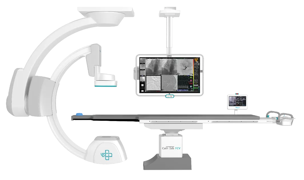
Потолочная ангиографическая система PROXIMA CS
Потолочная ангиографическая система PROXIMA CS
Proxima CS — это сочетание оптимального качества изображения при низкой дозе рентгеновского облучения с универсальными и быстрыми движениями до 25 градусов в секунду. Позволяет пользователю выполнять сложные кардиологические и интервенционные процедуры.
Дополнительные сведения
| Производитель | |
|---|---|
| Страна Производитель | Индия |
Основные характеристики
● Передовая технология визуализации: оснащена технологией цифровой плоской панели для превосходного разрешения и четкости изображения.
● Гибкое позиционирование: система может похвастаться легким доступом к пациенту и несколькими вариантами позиционирования, что повышает эффективность рабочего процесса.
● Высокоскоростная визуализация: обеспечивает быстрые последовательности изображений, необходимые для захвата динамических сосудистых структур.
● Повышенная безопасность: интегрированные функции управления дозой обеспечивают оптимизированное воздействие радиации, уделяя первостепенное внимание безопасности пациента.
Применение
Proxima CS идеально подходит для широкого спектра сосудистых и интервенционных процедур, включая:
● Периферическая ангиография
● Нейроваскулярная визуализация
● Катетеризация сердца
● Электрофизиологические исследования
● Эндоваскулярное лечение
Преимущества
● Ротационная ангиография (РА) — это метод получения диагностических изображений во время движения гентри Cathlab.
● Доступно многомерное изображение с более подробной информацией об анатомии.
● Данные изображения RA можно использовать для создания трехмерного реконструированного объема сосудистой анатомии.
Эти реконструированные данные можно использовать для планирования вмешательства и лечения.
● RA помогает уменьшить объем используемого контрастного вещества, а также дозу облучения.
● RA также полезен в случае визуализации сложных сосудистых структур.
Особенности:
● Один широкоэкранный монитор с несколькими экранами (до 6).
● Отображение уровней дозы и предупреждений на экране монитора.
● Без коллимации излучения.
● Бесконтактные датчики предотвращения столкновений.
● Новое поколение программного обеспечения “SYNERGY ACQUISITION” для визуализации с низкой дозой.
● Возможность расширения до ротационного и трехмерного ангиосканирования.
● Включена функция онлайн-DSA.
● Эффективность СТЕНТА
Основные характеристики: Размер детектора 30 x 30 см. ● Разрешение 1,5K x 1,5K, DQE 80%. ● Высокое разрешение 55*, яркий цветной монитор с несколькими вариантами отображения / монитор медицинского класса. ● Визуализация без искажений. ● Моторизованное вращение и движение детектора вверх / вниз. ● Двойной инверторный генератор мощностью 100 кВт. ● Жидкометаллическая трубка для самых жестких клинических требований. ● Онлайн-цифровая субтракционная ангиография. ● Протоколы ASSURE для защиты от радиации. ● Функция просмотра стента и функция постепенного появления и исчезновения стента. ● Более длинный стол катетеризатора для охвата от головы до ног. ● Интеграция / взаимодействие FFR, IVUS и OCT.
ПРОГРАММНОЕ ОБЕСПЕЧЕНИЕ SYNERGY
Программное обеспечение для визуализации:
● Получение изображений до 30 кадров в секунду при разрешении 1,5K x 1,5K в флуороскопии и киносъемке.
● Хранилище флуороскопии.
● Улучшение контуров в реальном времени, отображение позитивных/негативных изображений, регулировка уровня окна, контрастность/яркость, электронный коллиматор, вертикальное и горизонтальное переворачивание изображения, вращение изображения, масштабирование с функцией панорамирования.
● Измерения — длина, угол, стеноз и площадь.
● QCA/QLVA.
● Эффективный рабочий процесс цифровой визуализации (DICOM 3.0).
● Сеть передачи изображений, совместимая с DICOM 3.0.
● CD/DVD-рекордер пациента DICOM. Готовность к печати DICOM/бумаги.
● Онлайн-DSA в реальном времени, дорожная карта, сдвиг пикселей, повторное маскирование и успокоение пиков (опционально).
● Улучшенное программное обеспечение для просмотра просвета и сохранения флюорографии.
Основные характеристики
● Передовая технология визуализации: оснащена технологией цифровой плоской панели для превосходного разрешения и четкости изображения.
● Гибкое позиционирование: система может похвастаться легким доступом к пациенту и несколькими вариантами позиционирования, что повышает эффективность рабочего процесса.
● Высокоскоростная визуализация: обеспечивает быстрые последовательности изображений, необходимые для захвата динамических сосудистых структур.
● Повышенная безопасность: интегрированные функции управления дозой обеспечивают оптимизированное воздействие радиации, уделяя первостепенное внимание безопасности пациента.
Применение
Proxima CS идеально подходит для широкого спектра сосудистых и интервенционных процедур, включая:
● Периферическая ангиография
● Нейроваскулярная визуализация
● Катетеризация сердца
● Электрофизиологические исследования
● Эндоваскулярное лечение
Преимущества
● Ротационная ангиография (РА) — это метод получения диагностических изображений во время движения гентри Cathlab.
● Доступно многомерное изображение с более подробной информацией об анатомии.
● Данные изображения RA можно использовать для создания трехмерного реконструированного объема сосудистой анатомии.
Эти реконструированные данные можно использовать для планирования вмешательства и лечения.
● RA помогает уменьшить объем используемого контрастного вещества, а также дозу облучения.
● RA также полезен в случае визуализации сложных сосудистых структур.
Особенности:
● Один широкоэкранный монитор с несколькими экранами (до 6).
● Отображение уровней дозы и предупреждений на экране монитора.
● Без коллимации излучения.
● Бесконтактные датчики предотвращения столкновений.
● Новое поколение программного обеспечения “SYNERGY ACQUISITION” для визуализации с низкой дозой.
● Возможность расширения до ротационного и трехмерного ангиосканирования.
● Включена функция онлайн-DSA.
● Эффективность СТЕНТА
No technical specifications available.

